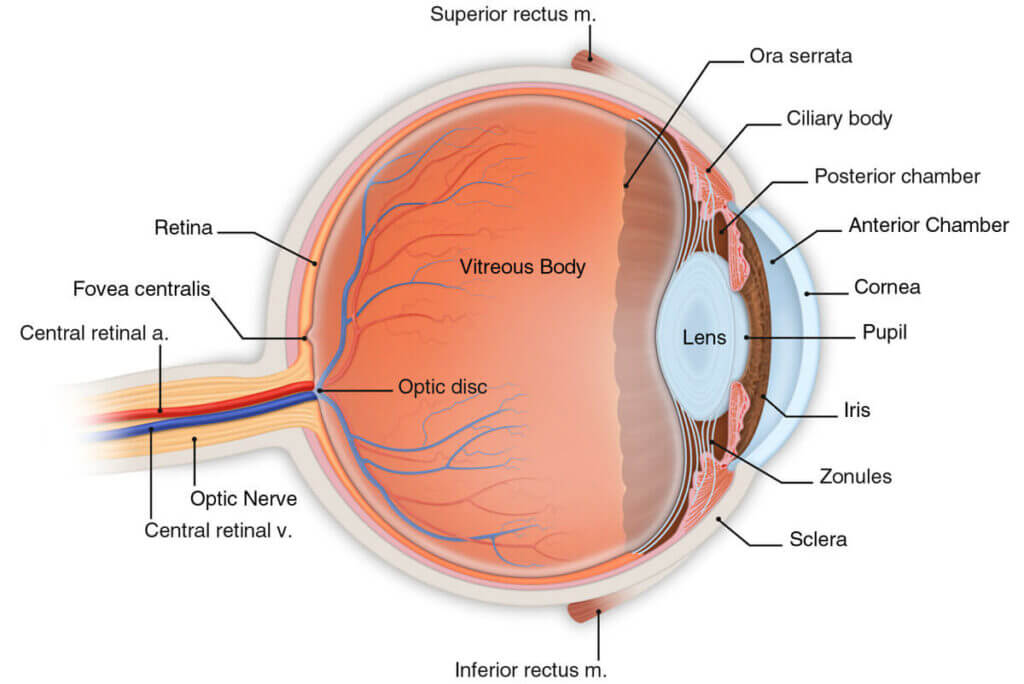
How the Eye Works

To understand the nature of vision problems, and how LASIK/PRK can correct them, you must first understand how the eye works. Although our eyes are very complex and almost magical, the process of how they work is quite simple to understand.
Light reflects off of objects and into our eyes. This light passes through the cornea, pupil and lens and is focused on the retina. After light passes through the pupil, the focus is fine tuned by the lens. This is called accommodation. The image focused onto the retina is transmitted to the brain through the optic nerve. The brain then interprets the image. When the brain receives and interprets the image, we have sight.
Cornea
The cornea is the clear outer part of the eye. Some people think of it like the windshield of the eye. It bends the light as it passes through; this bending is called refracting. Two-thirds of focusing occurs as the light is refracted (bent) by the cornea.
Iris and pupil
The iris is the colored part of your eye. It contains the muscles that open and close the pupil which lets light pass in. In low light the pupil dilates, opening to let in as much light as possible. In bright light, the pupil contracts, narrowing down to a very tiny opening and reducing the amount of light that passes through much like a shutter in a camera.
Lens
The lens is located just behind the pupil. After light passes through the pupil, the focus is fine tuned by the lens. Like the lens of a camera, the lens of the eye changes focus between near and far objects. This is called accommodation. Accommodation, or the focusing power of the eye, weakens with age. This weakening of accommodation is called presbyopia and is responsible for the need for reading glasses or bifocal lenses after the age of 44 for most people.
Retina
The retina is on the inside wall of the back of the eye. Some people consider the retina to be like the film in a camera. The retina contains photoreceptor cells. When light is focused on these cells they send the information to the brain through the optic nerve. The center of the retina, called the macula, is responsible for detecting fine details and color.
Optic Nerve
The optic nerve is the largest nerve in the body. It carries the information from the retina to the brain in order to interpret the image formed by the retina. The optic nerve is similar to a fiberoptic cable that transmits information from the retina to the brain.
Nearsighted (myopia)
When the cornea is curved too much or the eyeball is too long, light focuses in front of the retina, and has spread back out by the time it reaches the retina. This makes things off in the distance blurry.
Farsighted (hyperopia)
When the cornea is too flat or the eyeball is too short, the focal point is behind the retina, so the light is still spread out when it reaches the retina. You can see things clearly that are far away but cannot focus on objects near to you.
Astigmatism
This condition occurs when the eye is shaped like a football rather than a basketball and creates multiple focal points, instead of one, making the image blurry.
Presbyopia
Most people develop presbyopia in their 40’s. When people begin to develop presbyopia, they often think they are becoming farsighted because they have to hold things out away from their face to see them clearly, but it is not the same thing. It does not affect the shape of your cornea or the length of the eye. In presbyopia, the lens of your eye is no longer able to accommodate or make the fine adjustments needed for reading and fine work. Refractive surgery does not adjust the lens of the eye, but monovision can be used to eliminate the need for bifocals or reading glasses. Monovision can be done with PRK or LASIK.
