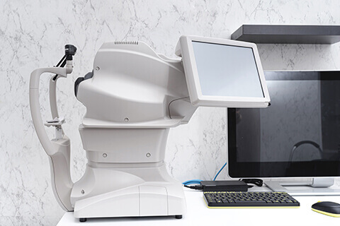
Diagnostic Technology
Western Laser Eye Associates has a number of diagnostic instruments in the clinic that are used for detection of a large number of eye conditions as well as to assess for PRK.
Our diagnostic technology includes:
Pentacam Corneal Mapping
The Pentacam is one of the most advanced technologies for corneal topography. Corneal topography mapping shows elevations and depressions in the corneal surface similar to the topography of a mountain range but on a micron scale. The Pentacam maps produce imaging and calculation of corneal curvature, contour, and tissue density at the microscopic level. This precise form of cornea mapping enables the accurate identification of PRK surgery candidates by measuring unique corneal tissue characteristics, such as posterior surface curvature and corneal thickness. The Pentacam map also includes a sophisticated analysis of each individual map that can be used to identify and study cases of corneal dystrophy such as keratoconus or pellucid corneal dystrophy.
Ocular Coherence Tomography (OCT)

OCT is a non-invasive imaging test that uses light waves to take cross-section pictures of your retina or optic nerve. The retina is the light-sensitive tissue lining the back of the eye and with OCT, each of the retina’s distinctive layers can be seen, allowing your ophthalmologist (Eye M.D.) to map and measure their thickness. These measurements help with early detection, diagnosis and treatment guidance for retinal diseases and conditions, including age-related macular degeneration and, diabetic eye disease, among others.
The OCT can also measure the optic nerve rim thickness in order to detect signs of glaucoma or optic nerve swelling. The optic nerve is the largest nerve in the body and is responsible for connecting the information received by the retina to the brain for proper visual functioning. Early detection of optic nerve changes can allow for treatment of eye diseases to prevent vision loss.
Humphrey Visual Field Analyzer
The Humphrey Visual Field Analyzer is considered the gold standard for the evaluation of peripheral (side) vision. There are a number of conditions including glaucoma, MS, stroke, and other central nervous system disorders that cause peripheral vision changes. Most people will not notice peripheral vision changes until they are quite advanced because they will naturally compensate by turning their head and in some cases if only one eye is affected they may not notice a change unless they cover one eye. With the Humphrey Visual Field Analyzer, conditions affecting the peripheral vision can be diagnosed and followed over time to recommend effective treatments.
The Humphrey Field Analyzer is also often used for driver vision evaluations in order to be sure the side vision is within safe standards as required by the provincial government.
Baily Lovie Contrast Sensitivity Test
Contrast sensitivity is the ability to discriminate between shades of grey. A standard eye chart is a high contrast and does not measure contrast sensitivity. Some people may require specialized contrast sensitivity testing as an occupational requirement. This test is also sometimes used in the diagnosis of eye conditions. This test must be administered by a trained optometrist under specific lighting conditions.
Orthoptics Testing
Orthoptics testing involves a series of specialized eye tests to determine how the eyes are working together. An optometrist with training in orthoptics does this specialized testing. Children with an eye turn or amblyopia (lazy eye) may need this testing in order to prescribe treatments to improve visual acuity or to avoid a worsening of vision. Orthoptics testing is also used for the diagnosis of eye movement disorders that may result from conditions such as stroke in older patients with difficulty keeping the eyes in balance while reading or doing other activities. Orthoptics testing is usually recommended by an ophthalmologist or optometrist and may need to be repeated over time in order to monitor for changes.
